Locality
From Hollick (1930) (p. 54)
"Anchorage Bay, opposite Northwestern Fishery Co.'s cannery, Chignik Bay, Alaska Peninsula (original No. 48), collected by W. W. Atwood and H. M. Eakin in 1908 (lot 5294) (pl. 17, fig. 1). Chignik Bay, 2 miles northeast of Alaska Packers Association cannery, Alaska Peninsula (original No. 958); collected by T. W. Stanton in 1904 (lot 3521) (pl. 17,
fig. 3). Yukon River, north bank, about 10 miles below Melozi telegraph station (original No. 18); collected by W. W. Atwood and H. M. Eakin in 1901 (lot 4633)." (pl. 17, fig. 2)
Description
From Hollick (1930) (p. 54)
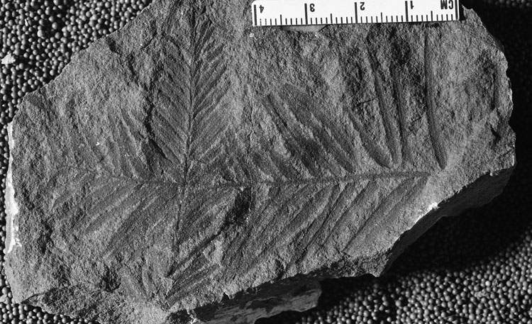
"Leafy twigs; leaves intermediate in shape and dimensions between Cephalotaxopsis magnifolia successiva, the variety last described, and Cephalotaxopsis microphilla laxa, the variety next described, being prevailingly smaller than the former and larger and
more rigid in habit than the latter."
Remarks
From Hollick (1930) (p. 54)
"The specimens included under this specific name represent one of the group forms of foliage referred to the genus Cephalotaxopsis that is very difficult to differentiate
satisfactorily from other similar group forms contained in the collections from Alaska.
It is inevitable that there should be considerable difference in the size of the leaves and in their disposition and arrangement in connection with the rachis to which they are attached, according to the part that happens to be preserved in any particular specimen, und hence dismembered specimens and detached leaves are often very diflicult to relegate to or to identify satisfactorily with anyone of the groups to which specific or varietal name has been given. The ultimate twig shown in Figure 1, for example, has short, closely approximated leaves and, if found isolated, would hardly be regarded as the same species as the larger twig or branch, with long, relatively widely spaced leaves, included in the same figure, or the median fragments shown in Figures 2 and 3."