Locality
Yukon-Koyukuk Basin Locality 11556
Description
Leaf: simple; symmetrical or asymmetrical; orbiculate; apex rounded; base lobate; margin dentate; teeth with rounded glandular apices, glands projecting beyond the apex, sides convex or concave, sinuses wide, shallow and rounded; margin entire at base; venation basal actinodromous, nine radiating veins; primary midvein weak to moderate, straight or slightly curved, occasionally sinuous, alternately branched along the apical half; a-pectinal veins strong, well developed forming an angle of 30-35° with the midvein at the base, straight or slightly recurved, becoming curved above, curvature often abrupt at points of departure of abmedial veins; a-abmedial veins moderate, curved, brochidodromous; b-pectinal veins strong, well developed, basally departing a-pectinals at angle of 30-35° curving by abrupt changes of course at points of departure of abmedial veins, curvature increasing near the margin to join the most basal a-abmedial at an acute or obtuse angle; b-abmedials moderate, slightly curved, curvature increasing abruptly at departure of veins which form medial veins of teeth, looping to join superadjacent b-abmedial at acute or obtuse angles, one or two tooth medial veins depart each loop; g-pectinal veins moderate to strong, departing the base at an angle of 30-35° to the b-pectinals, slightly curved except near margin where curvature abruptly increases at departure of tooth medial veins to join basal b-abmedial at an acute or obtuse angle; g-abmedials weak, curved, looping near margin to join superadjacent abmedial, one or two veins depart the loops abmedially to form medial veins of teeth; d-pectinals weak, forming angle of 30-35° to g-pectinal, curved, joining basal -abmedial, giving off brochidodromous weak d-abmedials; tertiary venation moderate to weak, orthogonal to random reticulate, often joining to form weak composite intersecondary veins; fourth and fifth order veins Ruding towards orthogonal, areolation moderately well developed, polygonal with a tendency to be quadrangular, veinlets usually branched once; ultimate marginal venation looped to incomplete.
Remarks
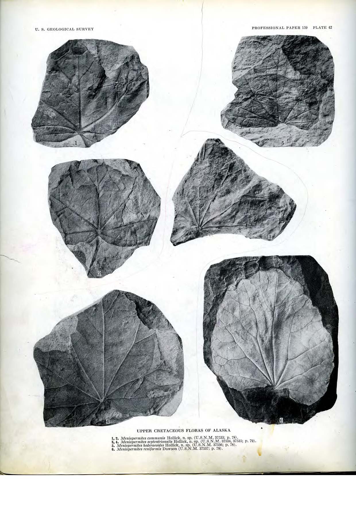
These specimens should be compared to the leaf Menispermites septentrionalis (Hollick, 1930, p. 79; Plate 42, Figs. 3, 4). The palmate radiating nervation is identical to that on the specimens illustrated by Hollick and matches his description, including the successive weakening of the "major lateral primaries" spreading away from the midvein. Hollick's specimens were incomplete and he had no knowledge of the leaf form, margin, or detailed venation. He described his specimens as being peltate but this condition is not seen in his illustrated specimens.
The teeth are particularly interesting in that they show characters of the Chloranthoid and Platanoid forms. In spite of good preservation no specimen shows the looping higher order laterals joining the medial vein. In all cases the higher order veins become extremely weak near the base of the apical gland.
The strong arching laterals, joining the medial vein at the base of the apical gland, and characteristic of the Chloranthoid tooth, are lacking. The failure of the laterals to join the medial vein and the successive brochidodromous looping of the laterals are, however, typical of Platanoid teeth. The medial vein in a Platanoid tooth usually attenuates towards the apex before gradually thickening and enclosing a cavity or foramen. In the fossil this cavity is invariably collapsed and is marked by a depression at the gland tip. With the exception of the glands of teeth near the base of specimen USGS 11556.29, this apical depression is not visible in these specimens. Other glands on specimen USGS 11556.29 show a pointed tip and on specimen USGS 11556.39 the gland is globular and there is no gradual thickening of the medial vein.
This form also occurs in the Cenomanian and Turonian of Russia.