Systematics
Tooth Types
In the course of reviewing the distribution of vegetative morphological characters throughout most sub-classes (based on Takhtajan, 1969) of extant dicotyledonous plants, Hickey and Wolfe (1975) presented a classification of tooth morphology. They recognized, described, and illustrated a number of tooth types of which, those relevant to the Arctic fossils illustrated in this catalogue are outlined as follows.
| Chloranthoid |
|
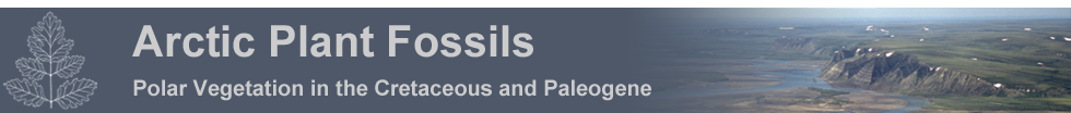
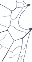
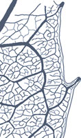
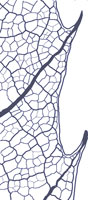
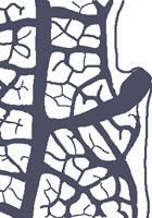
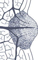
The Chloranthoid tooth is found in the Chloranthaceae and Illicales of the Magnoliidae, the Ranunculaceae, Glaucidiaceae, Hydrastidaceae, and Podophyllaceae of the Ranunculidae, the Trochodendrales, and possibly in a modified form in the Cercidiphyllales of the Hamamelididae. The Chloranthoid tooth typically has a clear, non-deciduous, swollen apical cap and varies in shape; acuminate-convex being common but acuminate-acuminate and concave-acuminate and concave-acuminate also occur. The venation of the tooth consists of a medial secondary or tertiary vein accompanied by two prominent, converging, higher order lateral veins which fuse with the medial vein at, or just below, the tooth apex. One of the converging laterals may be suppressed. |
| |
|
|
| |
|
 |
|
 |
|
 |
|
 |
|
 |
|
 |
| |
|
|
Monimioid
|
|
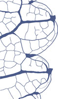
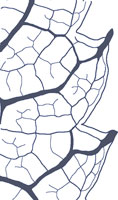
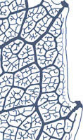
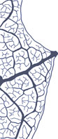
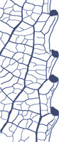
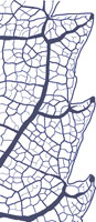
Monimioid teeth are found in the Monimiaceae and Trimeniaceae of the Magnoliidae. They have opaque non-glandular caps with an acute apex. The tooth shape is usually acuminate-convex and the venation consists of a secondary or tertiary vein entering the tooth medially and not joined by lateral veins. |
| |
|
|
| |
|
 |
|
 |
|
 |
|
 |
|
 |
|
|
| |
|
|
| Spinose |
|
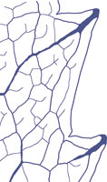
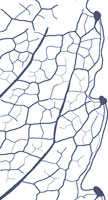
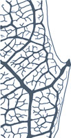
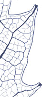
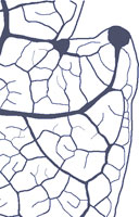
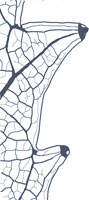
The Spinose tooth form has a wide distribution and has probably arisen a number of times. It is found in the Berberidaceae and Papaveraceae in the Ranunculidae, the Hamamelidaceae and some Fagales in the Hamamelididae, the Flacourtiaceae, the palmate dilleniids, and the Meliosmaceae in the Rosidae, where it could have arisen from the Cunonioid tooth type. The most obvious feature about this tooth is that the medial vein projects beyond the tooth apex. |
| |
|
|
| |
|
 |
|
|
|
|
|
|
|
|
|
|
| |
|
|
| Platanoid |
|
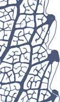
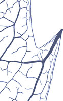
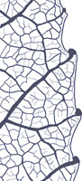
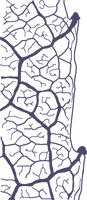
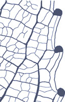
The Platanoid tooth is found in the Eupteleales, some Hamamelidales and Eucommiales of the Hamamelididae. It is characterized by having a medial secondary vein that becomes attenuated towards a glandular apex where it opens into a cavity or foramen. The medial vein is accompanied by higher order laterals forming a series of brochidodromous loops with the most apical pair converging on, but not reaching, the medial vein. |
| |
|
|
| |
|
 |
|
 |
|
|
|
|
|
|
|
|
| |
|
|
| Urticoid |
|
A somewhat similar tooth to the Platanoid is the Urticoid form found only in the Urticales of the Hamamelididae. This tooth type possesses higher order convergent lateral veins but has a non-glandular medial vein termination at or near the apex. |
| |
|
|
| |
|
 |
|
|
|
|
|
|
|
|
|
|
| |
|
|
| Dillenioid |
|
The pinnate and palmate dilleniid groups display a number of tooth types (Dillenioid, Theoid, Violoid, Salicoid, Malvoid, Cucurbitoid, Begonioid and Spinose) which are somewhat similar and may be derived from a common source. The Dillenioid and Theoid teeth are perhaps the more basic forms. Dillenioid teeth possess a medial principal vein which terminates in a clear glandular expanded apex with the vein often protruding beyond the tooth apex. |
| |
|
|
| |
|
|
|
|
|
|
|
|
|
|
|
|
| |
|
|
| Theoid |
|
The Theoid tooth exhibits an opaque glandular apical thickening of the medial vein capped by an opaque deciduous seta. |
| |
|
|
| |
|
 |
|
|
|
|
|
|
|
|
|
|
| |
|
|
| Violoid |
|
The Violoid tooth also has an opaque glandular apex but lacks the deciduous seta. |
| |
|
|
| |
|
 |
|
|
|
|
|
|
|
|
|
|
| |
|
|
| Salicoid |
|
The seta is retained in the Salicoid tooth but in a modified form as a dark, but not opaque, non-deciduous spherical callosity fused to the tooth apex. |
| |
|
|
| |
|
 |
|
 |
|
 |
|
|
|
|
|
|
| |
|
|
| Malvoid |
|
The Malvoid tooth is non-glandular but the vein principal veins runs directly to the tooth apex. |
| |
|
|
| |
|
|
|
|
|
|
|
|
|
|
|
|
| |
|
|
| Cucurbitoid |
|
Cucurbitoid and Begonioid teeth both have a translucent apical pad of densely packed cells, but differ in the strength and course of the subtending veins. |
| |
|
|
| |
|
 |
|
|
|
|
|
|
|
|
|
|
| |
|
|
| Begonioid |
|
Hickey and Wolfe (1975) inferred this to be a modification of the Cucurbitoid type in which one of the lateral veins appears to be strengthened at the expense of the medial and second lateral. |
| |
|
|
| |
|
|
|
|
|
|
|
|
|
|
|
|
| |
|
|
| Cunonioid |
|
Tooth with a small clear (in living forms) glandular apex, with the principal vein to the tooth branching below it, in or near the sinus and the sending one branch to the superadjacent secondary vein or to the sinus and the other branchh to the tooth apex on a deflected course along the apical side. |
| |
|
|
| |
|
 |
|
|
|
|
|
|
|
|
|
|
| |
|
|
| Rosoid |
|
Tooth with a large, clear (in living leaves) glandular apical opening (foramen) broadening distally from the sub-apical termination of the usually central principal vein of the tooth. A pair of lateral accessory veins of higher order (conjunctal veins) is connivant with the principal vein, follows a straight rather than a looped course, and terminates in the apical foramen. |
| |
|
|
| |
|
 |
|
|
|
|
|
|
|
|
|
|
| |
|
|
Success in correctly identifying tooth types from fossil material ultimately depends on the quality of preservation. The Chloranthoid, Monimioid, Platanoid, Urticoid, and Spinose teeth, all of which may be identified by means of tooth venation, gland geometry and morphology of the tooth, can probably be reliably distinguished from each other where preservation is reasonably good. The Dillenioid teeth, however, are characterized by gland opacity and the presence or absence of a seta or cap. Gland opacity is certain to change during fossilization and setae may be lost.
Cretaceous angiosperm leaf forms, particularly those in the late Early and early Late Cretaceous, exhibit considerable variability in venation and morphology. This variation is also likely to occur in the architecture of the tooth, perhaps even within a single leaf.
Hickey and Wolfe (1975) recognized that tooth types appear to be a stable and therefore useful systematic tool. With a few exceptions, notably the Spinose tooth which seems to have arisen more than once, tooth types are disjunctly distributed between the subclasses of extant dicotyledonous plants. This observation suggests modern tooth types, or their immediate precursors, may have arisen early in the evolution of the different subclasses and therefore might be a useful tool in the classification of fossil leaves. However, the various tooth types are recognized on the basis of an aggregation of characters. It is unlikely that all these characters were acquired simultaneously and it is reasonable to expect that the fossil record will show variants on these typical tooth forms. |
|